PDS logoPlanetary Data System
PDS Information
Find a Node - Use these links to navigate to any of the 8 publicly accessible PDS Nodes.

This bar indicates that you are within the PDS enterprise which includes 6 science discipline nodes and 2 support nodes which are overseen by the Project Management Office at NASA's Goddard Space Flight Center (GSFC). Each node is led by an expert in the subject discipline, supported by an advisory group of other practitioners of that discipline, and subject to selection and approval under a regular NASA Research Announcement.
Click here to return to the Photojournal Home Page Click here to view a list of Photojournal Image Galleries Photojournal_inner_header
Latest Images  |  Spacecraft & Technology  |  Animations  |  Space Images App  |  Feedback  |  Photojournal Search  

PIA16615: Calcium-Rich Veins in Martian Rocks
 Target Name:  Mars
 Is a satellite of:  Sol (our sun)
 Mission:  Mars Science Laboratory (MSL)
 Spacecraft:  Curiosity
 Instrument:  ChemCam
 Product Size:  3391 x 4134 pixels (w x h)
 Produced By:  LPGNantes 
 Full-Res TIFF:  PIA16615.tif (42.07 MB)
 Full-Res JPEG:  PIA16615.jpg (715 kB)

Click on the image above to download a moderately sized image in JPEG format (possibly reduced in size from original)

Original Caption Released with Image:


click here for larger view of figure 1 for PIA16615click here for larger view of figure 2 for PIA16615
Figure 1Figure 2
Click on an individual image for larger views

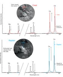
This graphic from NASA's Curiosity mission shows close-ups of light-toned veins in rocks in the "Yellowknife Bay" area of Mars together with analyses of their composition.

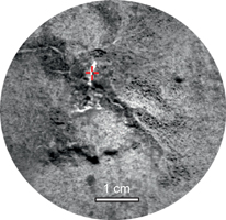
The top part of the image shows a close-up of the rock named "Crest," taken by the remote micro-imager (RMI) on Curiosity's Chemistry and Camera (ChemCam) instrument above the analysis of the elements detected by using ChemCam's laser to zap the target. The spectral profile of Crest's light-colored vein is shown in red, while that of a basaltic calibration target of known composition is shown in black.

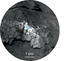
The bottom part of the image shows ChemCam's close-up of the rock named "Rapitan" with the analysis of its elemental composition. The spectral profile of Rapitan's light-colored vein is shown in blue, while that of a basaltic calibration target of known composition is shown in black.

These results suggest the veins are unlike typical basaltic material. They are depleted in silica and composed of a calcium-bearing mineral.

The ChemCam instrument took the RMI pictures and zapped lasers on Crest on Dec. 13, 2012, or the 125th sol, or Martian day, of operations. The ChemCam instrument took the RMI pictures and zapped lasers on Rapitan on Dec. 23, 2012, or the 135th sol, or Martian day, of operations.

Image Credit:
NASA/JPL-Caltech/LANL/CNES/IRAP/LPGNantes/CNRS

Image Addition Date:
2013-01-15